
Figure 2.
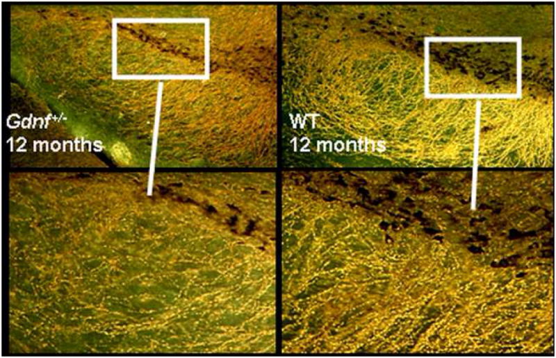
Darkfield illustration of the loss of TH-positive cell bodies and fibers in 12-month old Gdnf+/− mice (left side of panel) compared to age-matched WT controls (right side panel). Cell bodies in the Gdnf+/− mice are atrophied and a marked loss of TH-positive neurites in the pars reticulata can be seen.