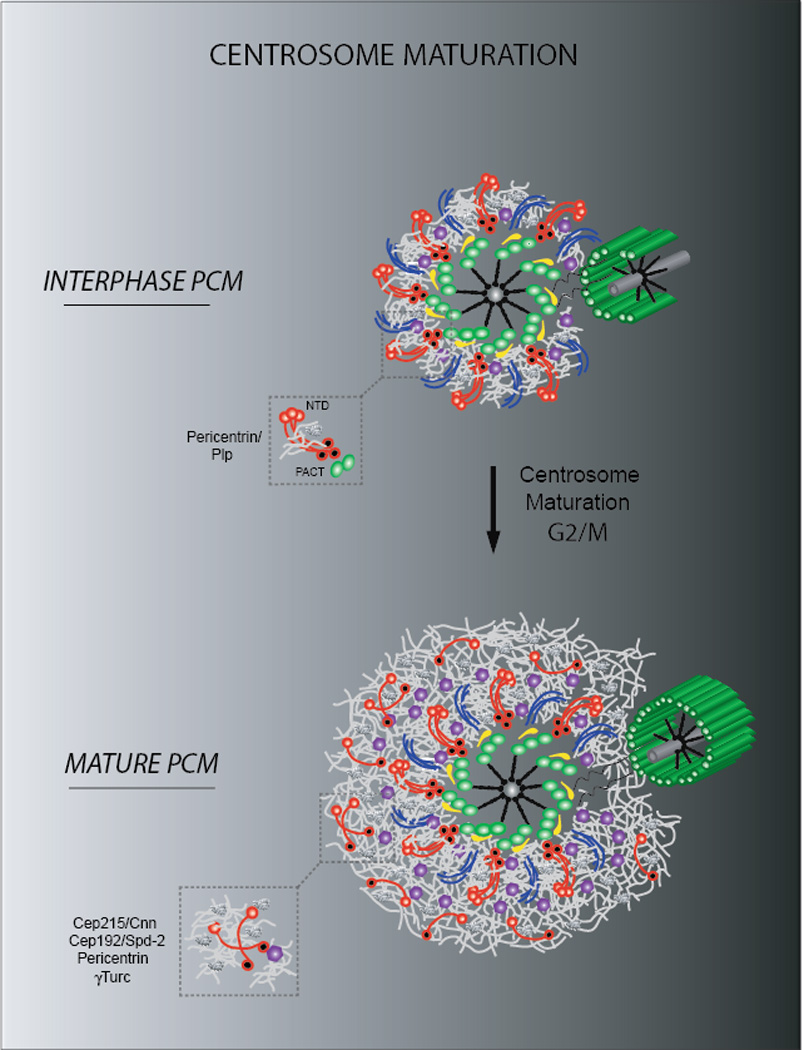
Figure 3. The PCM architecture during centrosome maturation.

Schematic representation of a mammalian centrosome during centrosome maturation. Note the expansion of the PCM proximal layer of interphase cells during G2/M through the formation of an outer matrix of Cep215, pericentrin and Cep192 molecules. γTuRC are embedded within the PCM and promote microtubule nucleation during mitosis. In contrast, Drosophila PCM expansion does not require the pericentrin homologue PLP.
