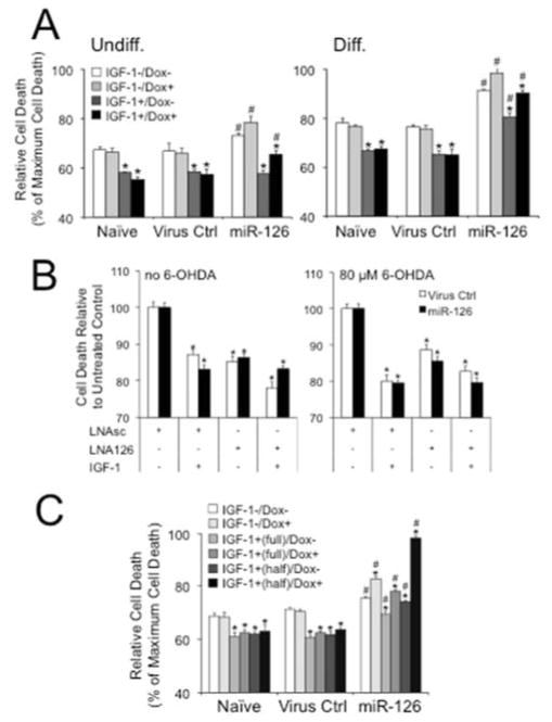
Figure 4.
Overexpression of miR-126 conveys vulnerability and its inhibition neuroprotection to 6-OHDA toxicity in neuroblastoma cells. (A) Treatment of undifferentiated (Undiff.) and differentiated (Diff.) SH-SY5Y cells with 80 μM 6-OHDA in the absence or presence of 20 ng/ml IGF-1 shows higher toxicity and less protective effects of the trophic factor in Dox+ (200 ng/ml) miR-126 transduced cells when compared to Naïve or Virus Ctrl. Results are from LDH assays and plotted as relative cell death to Triton-X (1%) induced maximum cell death (*P < 0.05 comparing IGF-1+ to IGF-1− condition; #P < 0.05 comparing miR-126 to Naïve or Virus Ctrl). (B) Inhibition of miR-126 increases cell survival of untreated and 6-OHDA treated SH-SY5Y cells. Dox-treated Virus Ctrl and miR-126 transduced cells were transfected with LNAsc or LNA126 and cultured in the absence or presence of 80 μM 6-OHDA and 20 ng/ml IGF-1. Data are plotted as percent cell death relative to untreated LNAsc controls (*P < 0.05 comparing IGF-1 and LNA126 to untreated LNAsc conditions). (C) Treatment of differentiated PC12 cells with 250 μM 6-OHDA in the absence or presence of 20 ng/ml IGF-1 shows higher toxicity and less protective effects of the trophic factor in Dox+ (200 ng/ml) miR-126 transduced cells when compared to Naïve or Virus Ctrl. Cells were differentiated according to a protocol by Chung et al., (Chung, et al., 2010), and a protective effect of IGF-1 was tested throughout (“full”) or only in the late phase (“half”) of differentiation (see Supplementary Fig. 5A) (*P < 0.05 comparing IGF-1+ to IGF-1− condition; #P < 0.05 comparing miR-126 to Naïve or Virus Ctrl).

