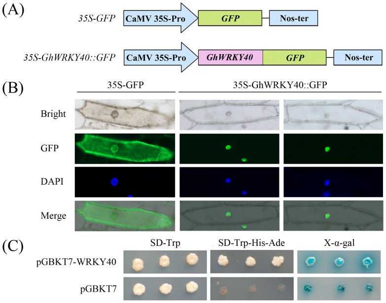
Figure 2. Subcellular localization of the GhWRKY40 protein and transcriptional activation of the GhWRKY40 gene.
(A) Schematic diagram of the 35S-GhWRKY40::GFP fusion protein construct and the 35S-GFP construct. (B) Transient expression of the 35S-GFP and 35S-GhWRKY40::GFP constructs in onion epidermal cells. Green fluorescence corresponding to the expressed proteins was observed with a fluorescence microscope 24 h after particle bombardment. The nuclei of the onion cells were visualized by DAPI staining. (C) Transactivation of the GhWRKY40 gene in yeast. The vector pGBKT7 was used as a control. The transformed yeast culture was streaked onto SD/-Trp or SD/-Trp-His-Ade medium, and the α-galactosidase activity was determined. Three independent experiments were performed.

