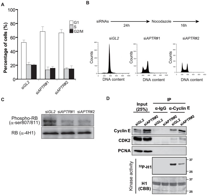
Figure 2. APTR depletion suppresses the G1/S phase progression.
(A) MCF10A cells transfected with siAPTR #1 or #2 accumulate in the G1 phase of the cell-cycle as measured by two color FACS for propidium-iodide and BrdU (mean ±s.e.m., n = 3). (B) 293T cells transfected with siGL2 or siAPTR #2 were analyzed by FACS for propidium-iodide in presence of Nocodazole (0.1 µg/ml). Schematic of the Nocodazole treatment procedure was presented on the top. (C) Immunoblot shows that Retinoblastoma protein is hypo-phosphorylated in 293T cells transfected with siAPTR #1 and #2. Total RB protein was analyzed as a loading control. (D) Reduced kinase activity of Cyclin E/CDK2 in 293T cells transfected with siAPTR#2. Cyclin E1 and CDK2 were analyzed by IP and immunoblot with indicated antibodies. PCNA was analyzed as a loading control. Kinase activity: autoradiogram of 32P labeled histone H1 after in vitro kinase assays with immunoprecipitates. CBB; coomassie blue staining to show equal amounts of H1 were added to all the lanes.

