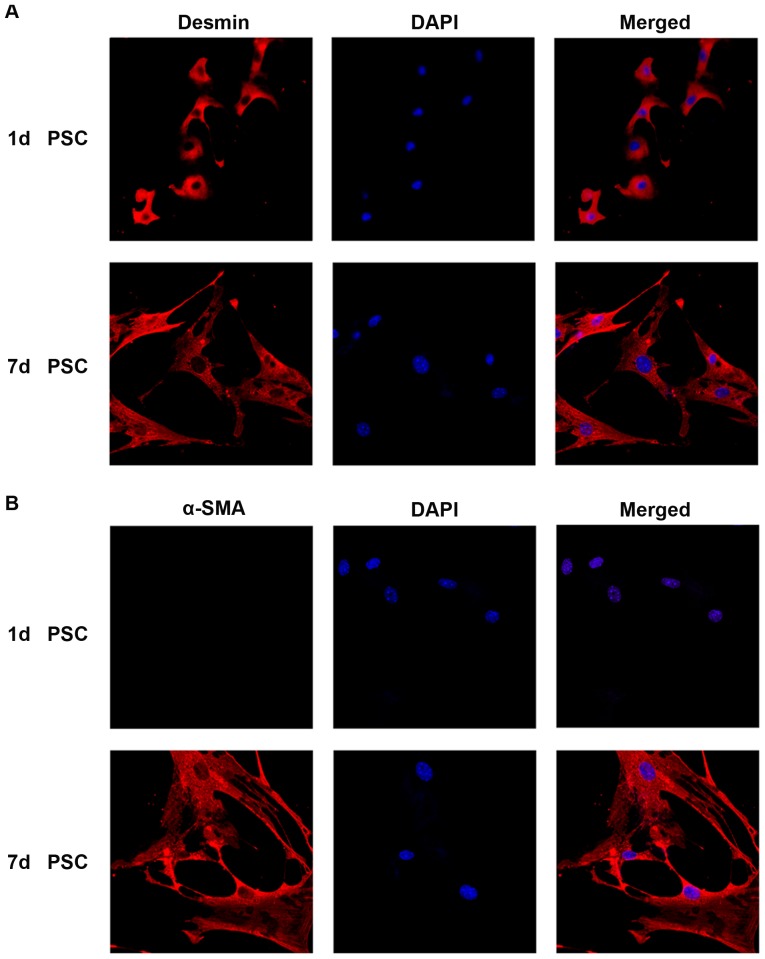
Figure 1. Mouse PSCs activation in vitro.
Immunofluorescence analysis of desmin and α-SMA during the activation of PSCs (A) Immunofluorescence staining of desmin in day 1 (quiescent) and day 7 (fully activated) PSCs. (B) Immunofluorescence staining of α-SMA in day 1 (quiescent) and day 7 (fully activated) PSCs. DAPI (blue) was used to counterstain nuclei (magnification ×400).

