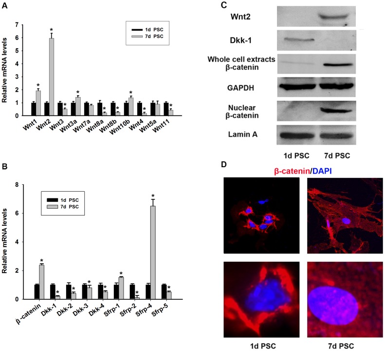
Figure 3. Expression of Wnt/β-catenin, Dkks and sFRPs families during activation of PSCs in vitro.
Quantitative RT-PCR detection of Wnt1, 2, 3, 3a, 4, 5a, 7a, 8a, 8b, 10b, 11 (A) and β-catenin and Dkk-1, 2, 3, 4 and sFRP-1, 2, 4, 5 (B) in quiescent and activated PSCs. Data are presented as mean±SD from three independent experiments. *p<0.05 compared with quiescent PSCs. (C) The protein levels of Wnt 2, Dkk-1 and β-catenin in quiescent and activated PSCs were detected by western blotting. (D) Immunofluorescent staining of β-catenin (red) in PSCs. DAPI (blue) was used to counterstain nuclei. Nuclear translocation of β-catenin in activated PSCs was observed.

