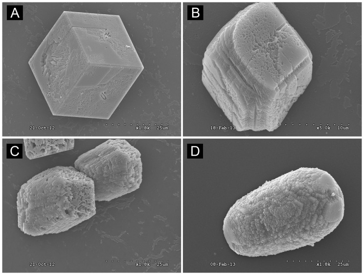
Figure 3. Scanning Electron Microscope Images of calcite modified by rhOtolin-1 alone or in combination with rmOC90.
Varying concentrations of rhOtolin-1 and rmOC90 protein, (A) 0 nM rhOtolin-1 and rmOC90; (B) 1000 nM rmOC90; (C) 667 nM rhOtolin-1; (D) 667 nM rhOtolin-1+500 nM rmOC90 were dissolved in 7.5 mM CaCl2 growth solution. Crystals were grown for 48 hours by slow evaporation of NH4HCO3 into growth solution. Crystals were examined with JEOL JSM 6320F Field Emission scanning electron microscopy. Scale Bar, A, C, and D: 25 µm; B: 10 µm.

