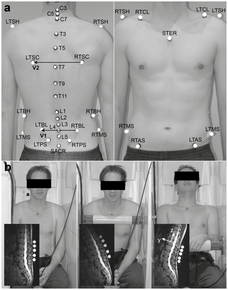
Figure 2. Measurement set-up including (a) the “IfB-marker-set” of the trunk and the pelvis (for explanation of abbreviations and for segmental allocation, see Table 2) and (b) the three analysed seating positions with an example of a corresponding MR image including a local coordinate system of a vertebral body.
Upright seating position: lower spine had partial contact with the backrest, and the whole upper body was in an upright position. Flexed seating position: upper body was tilted about 30° forward and supported on a bar, while the arms were rested on their lap. Extended seating position: the subject's bottom was pushed approximately 20 cm forward and the head was supported by the backrest. (from left to right).

