Fig. 7.
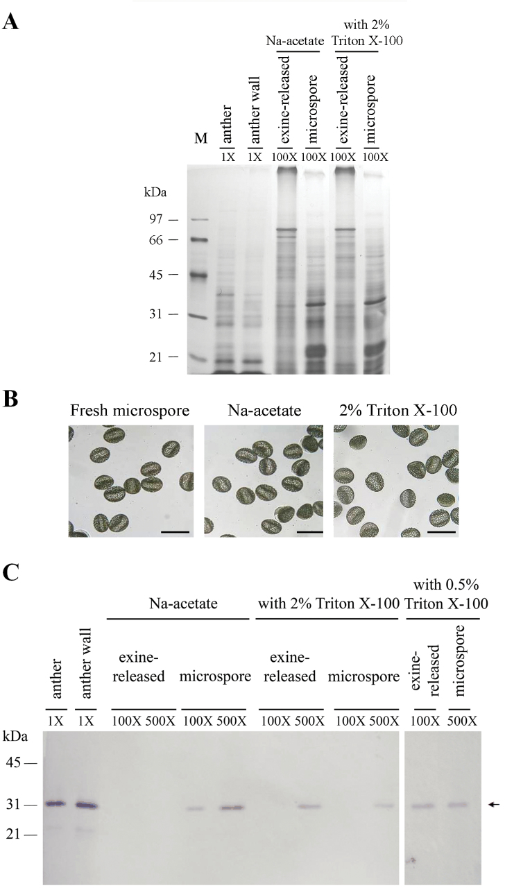
Distribution of the LLA1271 protein in fractions of distinct origins separated from anthers of L. longiflorum. SDS–PAGE of proteins in the anther of 34–46mm buds (total) and separated fractions including the anther wall, exine-released, and microspore fractions. Proteins were released from the exine layer of microspores by an aqueous solution of sodium acetate with or without the addition of either 0.5% or 2% Triton X-100. The gel was either stained with silver (A) or electroblotted onto nitrocellulose and immunologically detected using affinity-purified LLA1271 antibodies at a 1:20 dilution (C). Molecular mass markers in kDa are indicated on the left side. Different proportions of individual samples were applied to the lanes, and these proportions relative to an equal amount of the anther are shown in the gel. (B) Light microscopic photographs of fresh microspores, and microspores after treatment with an aqueous solution with or without 2% Triton X-100. The scale bar represents 100 μm.

