
Figure 1. Development of inducible stable clones and the ovarian orthotopic model.
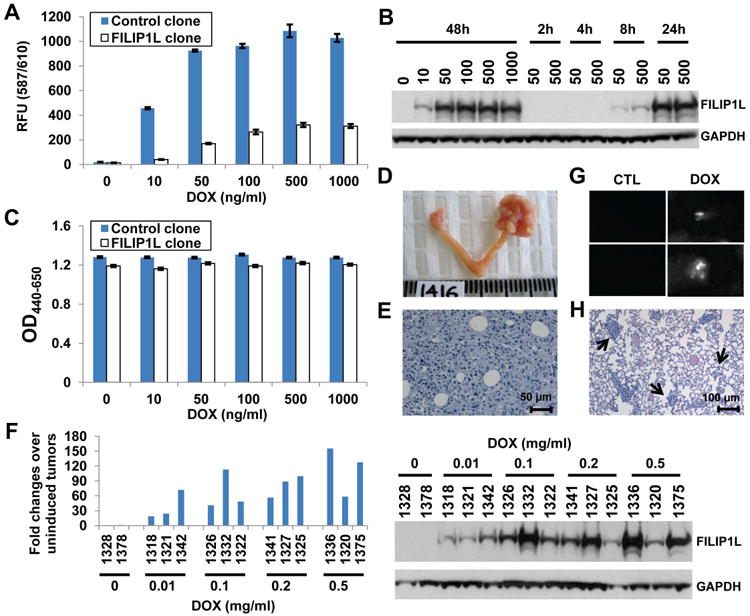
A, mCherry fluorescence measurement for control and FILIP1L clones treated with DOX for the indicated concentration. The y axis represents mCherry fluorescence measured at 587/610 nm (excitation/emission) (n=3). B, Immunoblot analysis for FILIP1L in FILIP1L clone treated with DOX for the indicated concentration (ng/ml) and time. C, WST1 cell proliferation assay for the same cells used in section A. The y axis represents cell proliferation measured with OD440-650nm (n=3). D, Ovarian orthotopic model. Mouse ovary from uninjected control (left) and FILIP1L clone-injected (right) 17 days after injection. E, H&E staining of ovarian orthotopic tumor shown in section D. F, qRT-PCR analysis for FILIP1L conducted on cDNA from FILIP1L clone-derived tumors (top). Drinking water containing the indicated concentration of DOX (mg/ml) was used to induce FILIP1L expression in mice. The numbers on the x axis indicate each mouse ID. The y axis represents fold change of each tumor over uninduced tumors (n=2), where each value was standardized with the housekeeping gene hRPL7. Immunoblot analysis for FILIP1L in the same tumor lysates used for qRT-PCR analysis (bottom). G, mCherry fluorescence of lungs from control clone-derived tumor-bearing mice 14 days after injection. Lungs were taken two days after giving mice drinking water containing 0.1 mg/ml DOX. White signals indicate lung metastases. H, H&E staining of the same lungs used in section G. Arrows indicate large (more than 50 cells)-, medium (6-50 cells)- and small (2-5 cells)-sized lung metastases, which were subjected to counting.