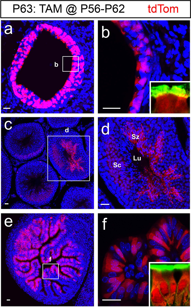
Figure 3. Foxj1 expression in peripheral organs.
Recombination induced in peripheral organs after 7 days of TAM administration beginning at P56 were analyzed 24 hours after the last TAM injection (P63: TAM@P56-P62). (a) Cross section of the lung epithelium showing tdTom+ cells in the primary bronchioles. (b) High magnification of primary bronchioles expressing tdTom in the boxed area. Inset reveals the tdTom+ cells as ciliated cells. (c) Cross section through the testis shows tdTom+ cells in the seminiferous tubule. (d) High magnification image of boxed area in (c) reveals the tdTom+ population in the seminiferous tubules includes the spermatocytes (Sc) as well as the spermatozoa (Sz) with elongated nucleus adjacent to the lumen (Lu). (e) Cross section of a female Foxj1CreERT2::GFP ampullary segment of the oviduct shows tdTom+ cells in the ciliated epithelium. (f) High magnification of boxed area in (e) highlights the tdTom+ ciliated cells in the epithelial folds. Insets in b and f show that tdTom+ cells are ciliated indicated by α-tubulin staining (green). Blue: DAPI nuclear staining in all images. Scale bars: 20µm in all images.

