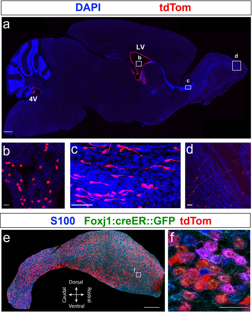
Figure 5. Labeling of the Foxj1 lineage in the postnatal forebrain.
(a) Sagittal section of a P21 brain from Foxj1CreERT2::GFP mice, induced with TAM at P0, shows tdTom+ cells (red) in the fourth ventricle (4V), lateral ventricle (LV), choroid plexus (b), rostral migratory stream (RMS; c), and olfactory bulbs (OB; d). (b) High magnification image of boxed area in (a) shows sporadic expression of tdTom+ cells in the choroid plexus of the LV. (c) High magnification of boxed area in (a) reveals numerous tdTom+ neuroblasts migrating in the RMS towards the OB, as reported in a previous study (Jacquet et al., 2011). (d) High magnification view of boxed area in (a) shows tdTom+ granule neurons in the OB. (e) S100 (blue) stained wholemount excised from the lateral ventricle from P21 Foxj1CreERT2::GFP mice brain reveals robust colocalization with tdTom+ ependymal cells. (f) High magnification image of boxed area in (e) reveals high degree of colocalization between S100 stained ependyma and tdTom+ cells. Scale bars: 500µm in (a) and (e); 50µm in (b), (c), and (d); 20µm in (f).

