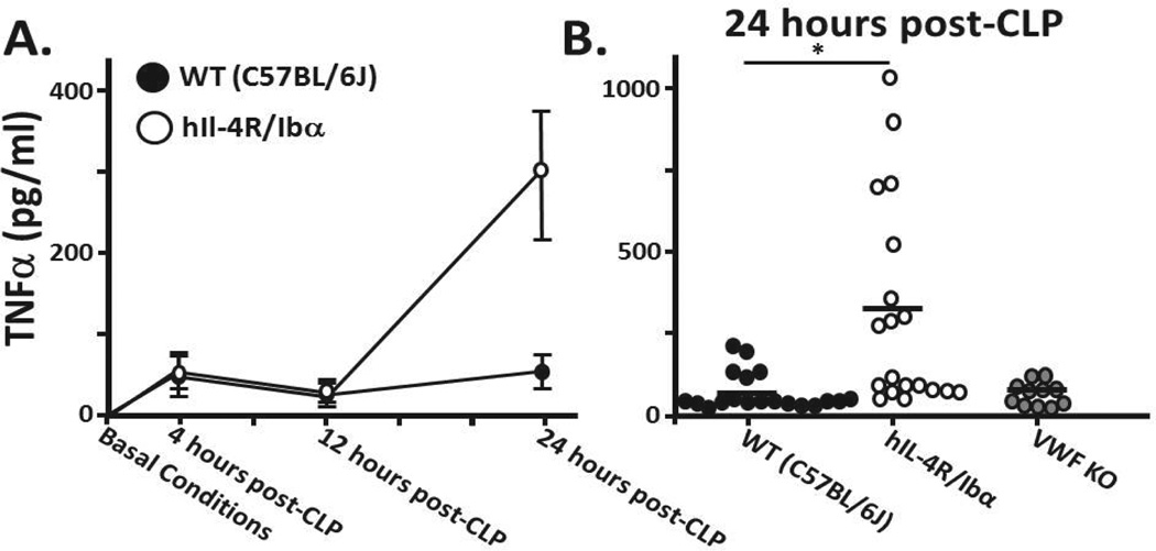
Figure 4. TNFα levels following CLP.
(A) Serum was collected from mice under basal conditions as well as 4, 12 and 24 hours post CLP. Under non-septic (normal) conditions, TNFα is undetectable in serum. At 4 and 12 hours wild type and hIL-4R/Ibα samples exhibit indistinguishable serum TNFα levels. A marked difference is observed at 24 hours following sepsis induction. (B) TNFα levels of individual mice 24 hours post-CLP show on average significantly elevated TNFα levels in those mice lacking GP Ib-IX expression (p = 0.005). Comparison of wild-type and VWF KO show no distinction between serum TNFαlevels (p = 0.933). A horizontal bar represents the overall mean. N = 19 (WT); N = 19 (hIL-4R/Ibα); N = 11 (VWF KO).

