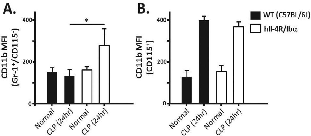
Figure 6. Mac-1 expression changes 24 hours post-CLP.
Blood isolated from wild-type (WT) and hIL-4R/Ibα mice under normal conditions or following CLP was stained using Gr-1 (neutrophils), CD115 (monocytes) and CD11b (Mac-1). Expression of Mac-1 for both the (A) neutrophil and (B) monocyte populations did not differ between strains in the absence of CLP (normal). Twenty four hrs following CLP, hIL-4R/Ibα display increased surface Mac-1 staining on the surface of neutrophils (p = 0.0348). Monocyte Mac-1 did not differ between strains 24 hours post-CLP. N = 6 for Normal samples and N = 11 for CLP samples.

