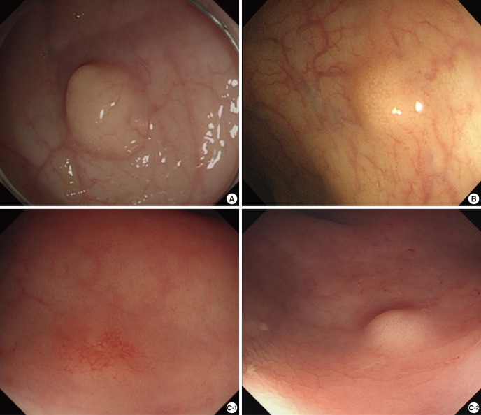
Fig. 1.
Color and endoscopic morphology of rectal carcinoid tumor. Among the 98 cases, tumors were yellow in 85 cases, white and pale in 10 cases, and the other three cases were similar in color to the surrounding normal mucosa. Tumor morphology after biopsy was classified into protruded type in 51 cases, slightly elevated or flat type in 43 and depressed or ulcerative type in 4. (A) The tumor is white and pale and morphologically belonged to the protruded type. (B) The color of tumor is yellow and it is of the slightly elevated type. (C) In the C-1 image, the tumor is a depressed type, and the color is neither yellow nor white. It could not be easily distinguished from the surrounding mucosa, but the tumor had definite capillary bed. C-2 image was the same patient's endoscopic image before the biopsy was taken. It shows typical features of carcinoid tumor, which was yellow-colored and slightly elevated type.

