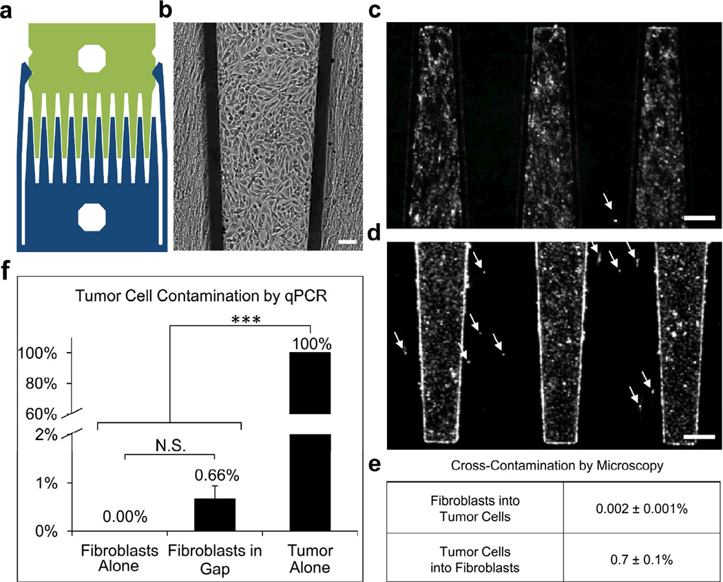
Figure 1.
Comb substrates allow cells to be cultured in close proximity with minimal cross-contamination. (a) Diagram of device with paired combs locked into the gap configuration. (b) Brightfield reflected light image of HT1080 tumor cells (cobblestone cells) and human lung fibroblasts (elongated cells) in gap co-culture after 48 h, scale bar 100 µm. (c–d) Fluorescent images with arrows highlighting (c) a DiI-labeled fibroblast contaminating the tumor population, and (d) DiI-labeled tumor cells contaminating the fibroblast population in gap co-culture, scale bar 250 µm. (e) Quantification of fluorescent images shows minimal cross-contamination. (f) Minimal tumor contamination of the fibroblast population in gap co-culture, as determined by qRT-PCR for the tumor cell marker, TERT, relative to HPRT. Samples displaying statistically significant changes (Student’s t-test) are indicated (*** = p < 0.005) and relevant changes that are not significant are denoted (N.S.). Error bars are SEM.

