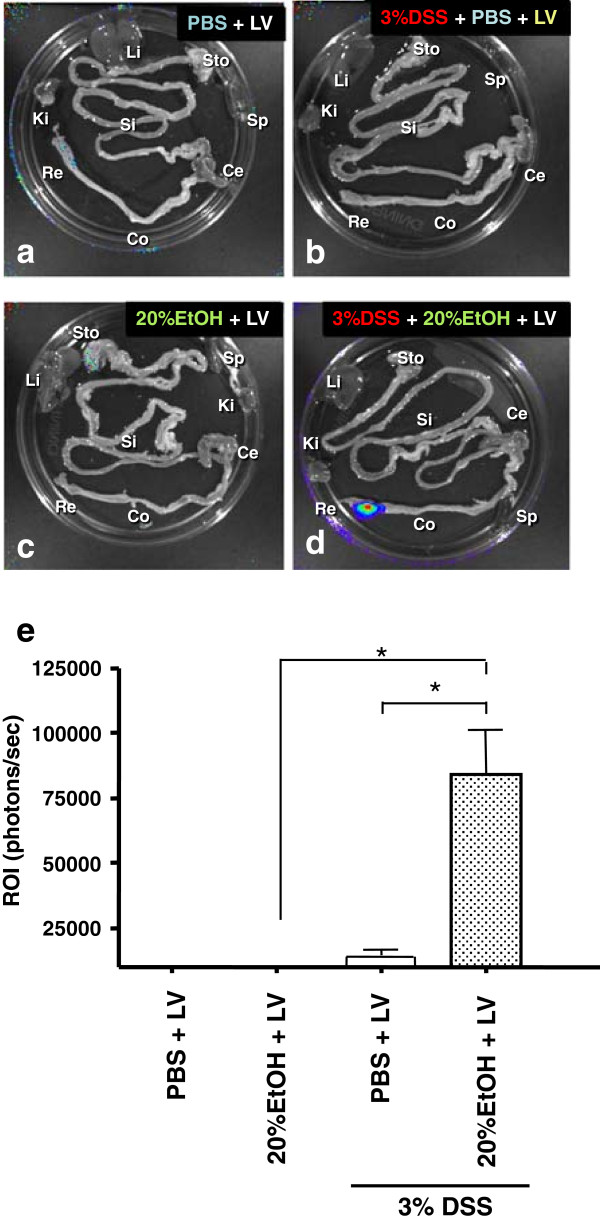
Figure 2.
Optimization of mucosal LV transfection. Ex vivo Bioluminescence imaging (BLI) analyses. Ex vivo BLI quantification and analyses of GI tract following intrarectal administration of LV expressing firefly luciferase (fLuc) on either colon with (a) PBS only, (b) with PBS followed by 3% DSS, (c) with 20% EtOH only, or (d) with 20% EtOH followed by 3% DSS. This pseudocolor image, superimposed on a gray scale reference image, uses color (blue: least intense; red: most intense) to illustrate signal strength. Abbreviations: Sto: stomach; Si: small intestine; Co: colon; Ce: Cecum; Re: rectum; Li: liver; Ki: kidney; Sp: spleen. (e) quantifies photon emission (p/s/cm2/sr) in the distal colon. *p < 0.05.

