
FIG. 5.
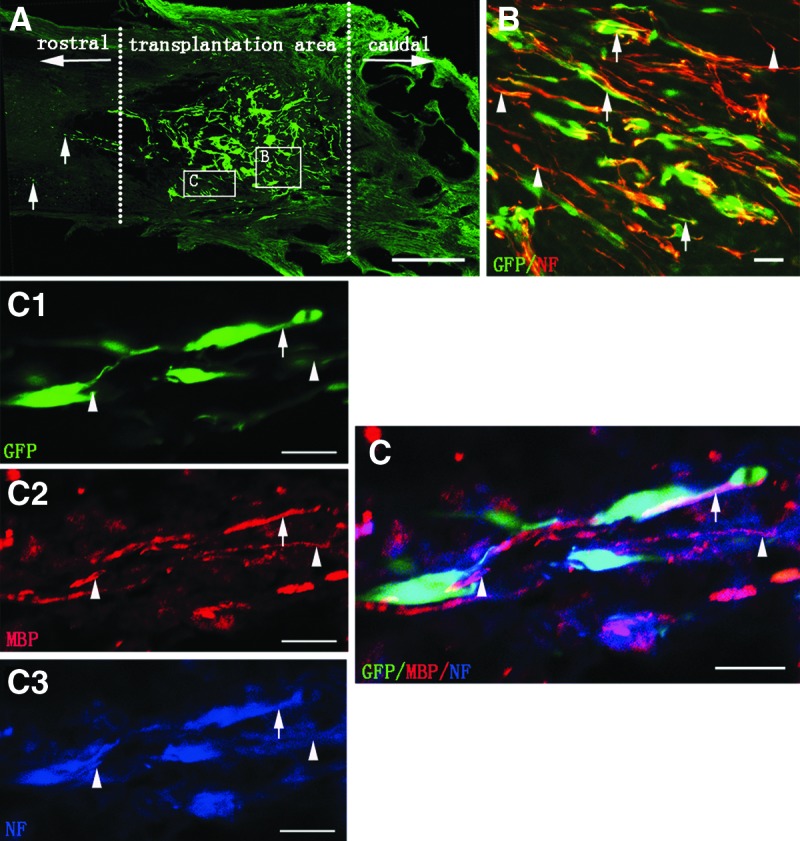
Cell survival, innervation, and myelination of the tissue-engineered neural scaffold transplanted in injured spinal cord. (A) Showing the cell survival of transplanted scaffold at eighth week in the N-SCs+T-NSCs group in a longitudinal section of injured spinal cord. Note that GFP-positive cells (green) of the neural scaffold transplanted are mainly distributed in the injury/transplantation area of spinal cord. The neural scaffold appears to integrate well with host tissue. Some of the GFP-positive cells (arrows) have migrated farther to the rostral of the injury/transplantation area. Leading diagram (A) depicts the cells' sampling sites in the spinal cord. (B) Showing transplanted NSC-derived neurons. NF-positive neurons (GFP-positive labeling) extend their processes (arrows). Some NF-positive axons from host (arrowheads) are distributed around GFP- and NF-positive double labeling axons. (C) Myelination of axons in the injury/transplantation area. Axons both from host (arrowheads in C3 and C) or NSC-derived neurons (arrows in C1, C3 and C) undergo robust myelination (arrows in C2 and C). GFP and NF positive double labeling cells (arrows in C) with thin axon are wrapped by MBP positive myelin sheath (arrow in C2). Scale bars=500 μm in (A); 20 μm in (B) and (C).