
Figure 5.
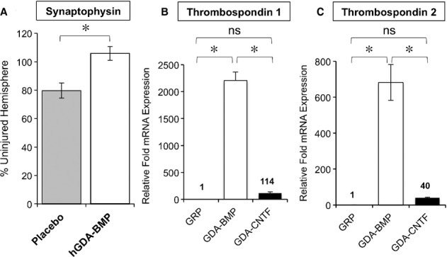
GDAsBMP express high levels of thrombospondin and increase synaptophysin expression in 6-OHDA-lesioned rats.
A Relative fluorescence intensity of synaptophysin staining in injured striatum of 6-OHDA injured animals after treatment with saline (placebo) or hGDAsBMP shows rescue to levels seen uninjured tissue by GDABMP transplantation. Expression was normalized to synaptophysin staining in contralateral, uninjured striatum. Mean of percentage ± s.d., n = 3. *P < 0.05, Unpaired t-test.
B, C Gene expression analysis of Thrombospondin 1 and 2 by semi-quantitative RT-PCR shows markedly higher levels of expression in GDAsBMP than in GRP cells or GDAsCNTF. Average relative expression normalized to expression found in glial precursors. Mean of n = 3 ± s.d., *P < 0.05 for all pairwise comparisons by One-Way ANOVA and Bonferroni Multiple Comparison Test except for comparison of GRP and GDACNTF conditions (ns).