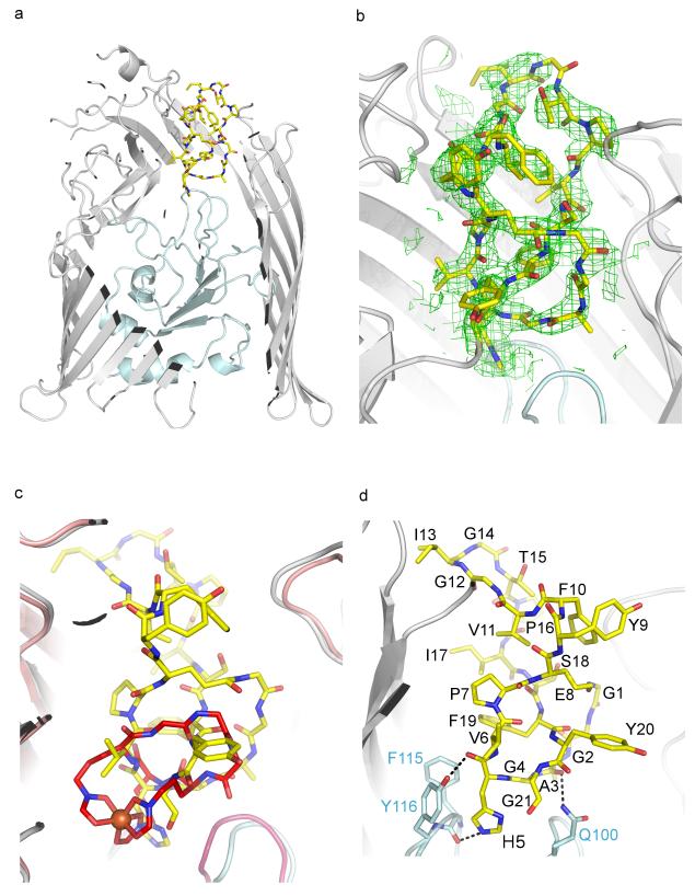
Fig 1.
Structure of E. coli FhuA in complex with MccJ25. (a) MccJ25 (yellow sticks) binds to the extracellular pocket of the outer membrane ferrichrome receptor FhuA. FhuA is displayed as grey ribbons and the plug domain is shown in light blue. The front face of FhuA, detergent molecules and LPS have been omitted for clarity. (b) Unambiguous 2Fo-Fc electron density was observed for MccJ25 after molecular replacement and rigid body refinement. MccJ25 was not included in the refinement but is shown for clarity. The map is contoured at 1σ (see also Supplementary Fig. 2). (c) MccJ25 (yellow sticks) mimics the binding of the ferrichrome (red sticks; iron is shown as an orange sphere). (d) MccJ25 displays three hydrogen bonds with the plug domain. Other interactions are shown in Supplementary Fig. 4. MccJ25 carbons are shown as yellow sticks, oxygens in red and nitrogen in blue. The FhuA plug domain side chains carbons are in light blue. The rest of the atoms are as in MccJ25.

