
Figure 3.
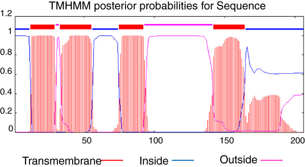
Protein profile analysis of the 22.91 kDa protein. The X-axis shows the amino acid position and the Y-axis indicates the probability of regions being located transmembrane (red), intracellular (blue) or extracellular (pink) predicted by TMHMM. The 11.5 kDa protein starts at the beginning of the second extracellular loop at amino acid position Y101.