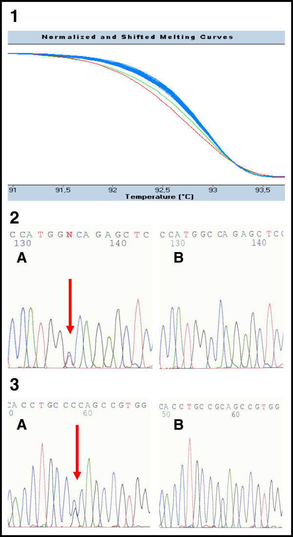
Figure 1.
The example of HRM analysis in patients 385 and 419 (exon 39). 1 Detection of mutations by HRM. The green curve in the diagram corresponds to the PCR fragment carrying the nonsense mutation c.11172G > A (p.Trp3724X) in patient 385. The red curve refers to the PCR fragment carrying the likely pathogenic substitution c.11248C > G (p.Arg3750Gly) in patient 419. The rest of fragments with blue curves are wild type samples (patients without sequence changes). 2, 3 Confirmation of mutations by direct sequencing in patient 385 (number 2) and patient 419 (number 3). Both sequences are in reverse direction. The letter A corresponds to the mutated sequence; B is the wild type.

