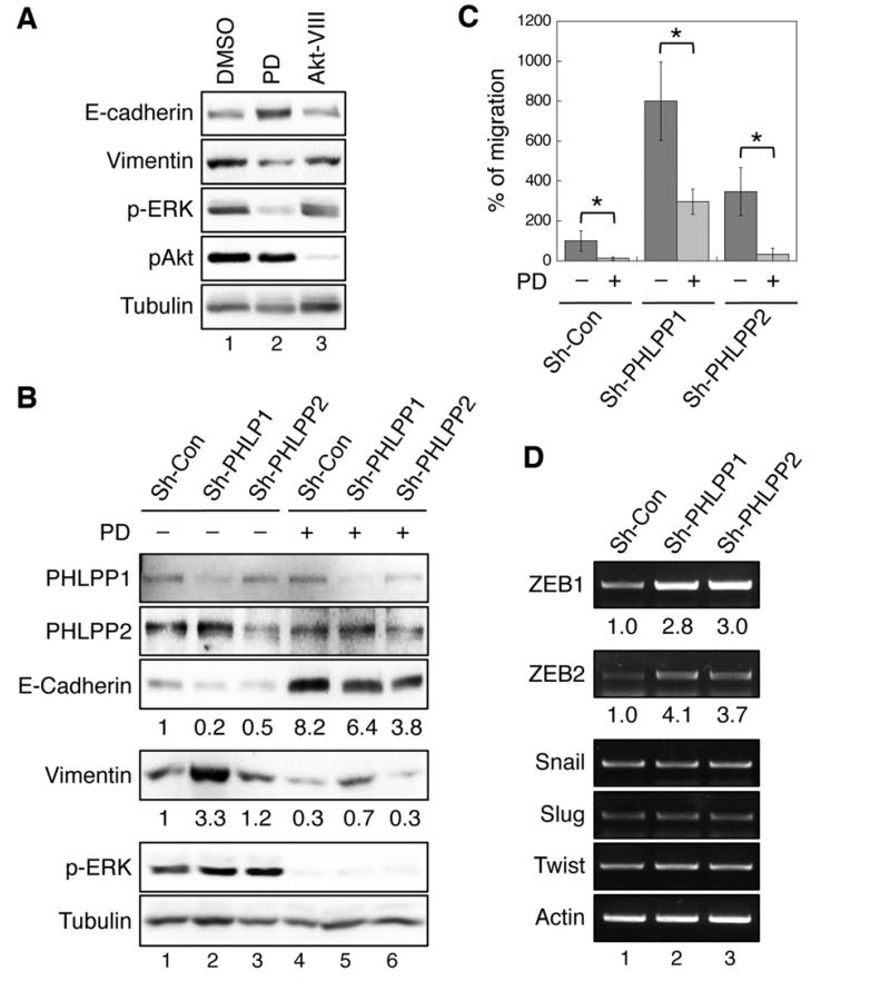
Figure 4. Loss of PHLPP promotes EMT by enhancing RAF/MEK/ERK signaling in CRC cells.
(A) SW480 cells were treated with DMSO, MEK inhibitor (PD0325901, 10 nM), or Akt-VIII (1 μM) for 30 hours, and cell lysates were analyzed by immunoblotting. (B) Stable SW480 knockdown cells were treated with DMSO or PD0325901 for 30 hours, and cell lysates were analyzed by immunoblotting. The relative expression of E-cadherine and vimentin were quantified by normalizing to tubulin. (C) Stable SW480 knockdown cells pretreated with DMSO or PD0325901 were subjected to Transwell migration assays in the presence or absence of PD0325901 using collagen and EGF as chemoattractants. Data shown in the graph represent the mean ± SD (n=3, * p<0.01 by two-sample t-tests). (F) The mRNA expression of ZEB1, ZEB2, Snail, Slug, Twist, and β-actin in control and PHLPP knockdown SW480 cells were assessed by semi-quantitative RT-PCR. The relative levels of ZEB1 and ZEB2 were quantified by normalizing to actin.

