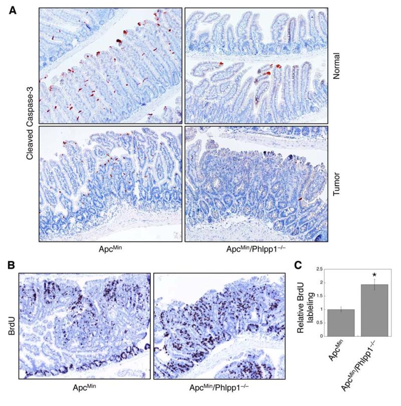
Figure 6. Phlpp1 deletion inhibits apoptosis and promotes cell proliferation in ApcMin mice.
(A) Intestine tissue sections from 24-week old ApcMin and ApcMin/Phlpp1−/− mice were stained for cleaved caspase 3. Representative images were taken from normal intestinal regions and adenomas. (B) Representative images showing BrdU labeled adenomas in ApcMin and ApcMin/Phlpp1−/− mice. (C) The numbers of BrdU positive cells in adenomas from ApcMin and ApcMin/Phlpp1−/− mice were quantified and analyzed using the Leica Application Suite EZ software. Ten randomly chosen tumors were averaged, and data in the graph represent the mean ± SD (* p<0.05).

