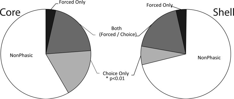
Figure 6.
The percentage of cells in the core (left) and shell (right) exhibiting nonphasic activity or patterned (phasic) cell firing during the preferred cues on force versus choice trials. There were similar percentages of cells in the core and shell that were phasic during only the force trials or during both choice and force trials. However, a significantly greater percentage of cue-related neurons were observed during choice trials in the core than in the shell.

