
Figure 3.
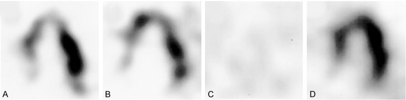
123/125I-MIBG SPECT long-axis views of the heart of Parkinson’s disease patients with a parkin mutation (A and B), idiopathic Parkinson’s disease (C), and a normal control (D). Reproduced with permission from [191] © John Wiley and Sons.
Official websites use .gov
A
.gov website belongs to an official
government organization in the United States.
Secure .gov websites use HTTPS
A lock (
) or https:// means you've safely
connected to the .gov website. Share sensitive
information only on official, secure websites.
123/125I-MIBG SPECT long-axis views of the heart of Parkinson’s disease patients with a parkin mutation (A and B), idiopathic Parkinson’s disease (C), and a normal control (D). Reproduced with permission from [191] © John Wiley and Sons.