
Figure 2.
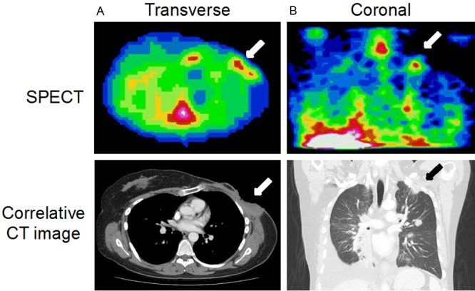
Representative images from two patients showing tumor accumulation of 111In-DTPA-hEGF with correlative CT images. A: Transverse SPECT image (Patient 13) acquired 24 h p.i. showing accumulation of 111In in an area of chest wall disease (arrow). B: Shows a coronal SPECT image (Patient 15), acquired 24 h p.i. demonstrating accumulation of radioactivity in a left infraclavicular fossa and lung apex tumor deposit.