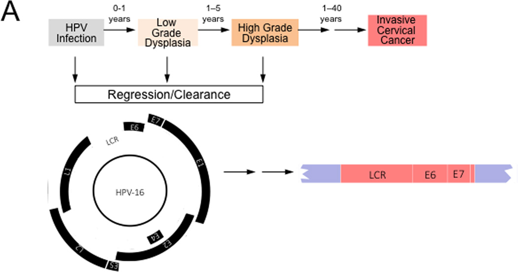
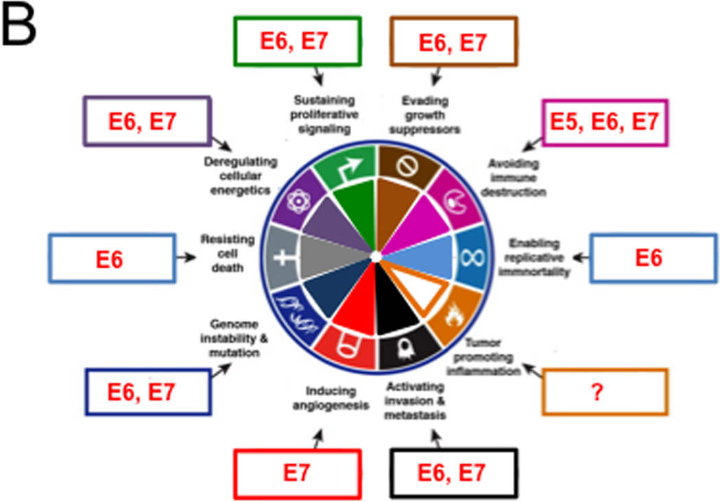
Figure 3. Hallmarks of Cancer analysis of HPV associated cervical carcinogenesis.
High-risk HPV infections can give rise to low-grade dysplasia (also referred to as cervical intraepithelial neoplasia 1 (CIN1)), which can progress to high-grade dysplasia (also referred to as CIN2/3). Many of these lesions spontaneously regress, presumably because of immune clearance by the host. These lesions contain episomal HPV genomes and expression of the viral genes is tightly controlled by the interplay of cellular and viral factors. Malignant progression to invasive cervical cancer is often a very slow process and cervical cancers can arise years or decades after the initial infection. Cervical carcinomas frequently contain integrated HPV sequences. Expression of the viral E2 transcriptional repressor is generally lost upon viral genome integration resulting in dysregulated viral gene expression from the viral Long Control Region (LCR). HPV E6 and E7 are consistently expressed even after genome integration and expression of these proteins is necessary for the maintenance of the transformed phenotype. See text for detail.
(B) High-risk HPV proteins target almost the entire spectrum of the Cancer Hallmarks. HPV genes that affect each hallmark are indicated within boxes around the wheel. See text for details


