
Fig. 1.
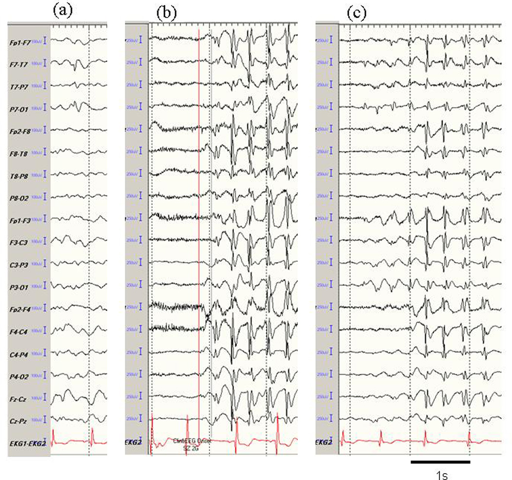
Representative EEG activity in Case 1. (a) single spike in the left temporal area, (b) synchronous generalized 3 Hz spike-and-wave complexes from seizure onset, and (c) secondarily generalized 3 Hz spike-and-wave complexes. After undergoing resection of a tumor in the left temporal lobe, the patient was seizure-free, and the epileptic activities shown here also disappeared completely.