
Fig. 3.
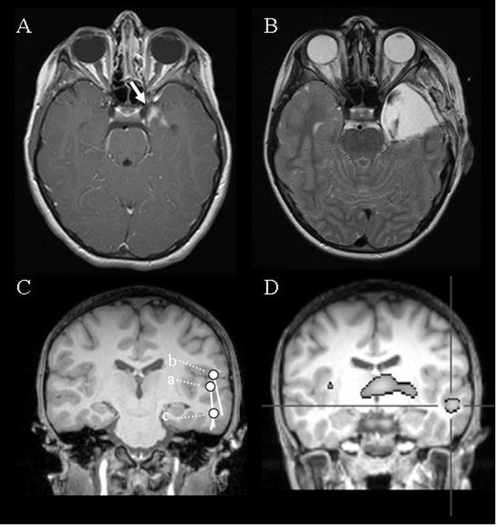
From top, EEG and MEG waveforms, magnified spike waveform, contour map of MEG, and dipoles on individual MRI of representative epileptic discharges are shown for Case 2. (a) single spike in the left temporal area, (b) synchronous generalized 3 Hz spike-and-wave complex from seizure onset, and (c) secondarily generalized 3 Hz spike-and-wave complex. MEG: The analyzed portions of the MEG waveform are indicated by the broken box. Note that MEG tends to show spikes with greater clarity than EEG. The time latency between the left and right epileptic discharges during generalized spikes, seen in panel b, is 7 ms. Contour maps: Arrows on the contour maps indicate the estimated dipoles. The solid line indicates magnetic field efflux, and the broken line indicates magnetic field influx from the brain surface. MRI: The circle and bar shown on the MRI scans indicate the dipole location and orientation, respectively.