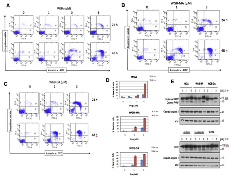
Fig 3.
Novel withanolides induce apoptosis in MTC cells. Dot-blot flow cytometry data for co-staining with Annexin V and PI after treatment with different concentrations of 3 novel withanolides, (A) WGA, (B) WGB-MA, and (C) WGB-DA. Significant apoptosis was observed at 6 μmol/L WGA, and 1 μmol/L WGB-MA, and WGB-DA. D, Quantitative apoptosis of MTC cells treated with these withanolides for 24 and 48 hours. More than 80% of cells were gated toward apoptotic cell death with only 3 μmol/L exposure of WGB-DA. E, Confirmation of apoptosis by Western Blot analysis, demonstrating activation of caspase 3 and cleavage of PARP in a dose-dependent manner, starting at 1 μmol/L of withanolide drug. This effect is less robust with the TKI drug compounds at similar concentrations. (Color version of figure is available online.)

