Fig. 4.

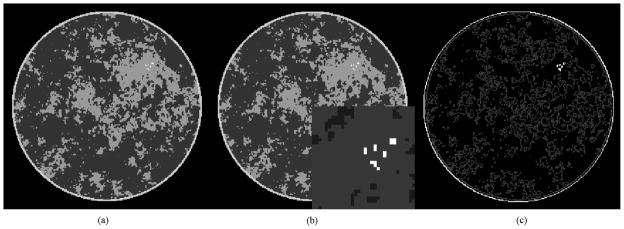
Left: 256 × 256 pixelized breast CT phantom used in the present study. Middle: same with ROI around microcalcifications shown magnified as inset. Right: the gradient magnitude image, which has a sparsity of 10019 nonzero pixel values.

Left: 256 × 256 pixelized breast CT phantom used in the present study. Middle: same with ROI around microcalcifications shown magnified as inset. Right: the gradient magnitude image, which has a sparsity of 10019 nonzero pixel values.