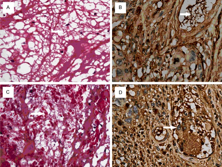
Figure 5.

Masson staining of mesh-like structure. Arrow: Mesh-like structure was nest-like biological filter (A, × 400, Masson staining), in which red blood cell dominant blood cells filled (C, × 400, Masson staining). In colon cancer, massive mesh-like structure (anti-fibrinogen antibody, 1: 100, × 400) was observed in venules, and cancer cells were also observed in this mesh-like structure (anti-fibrinogen antibody, 1: 100, D, × 400).
