Figure 1.
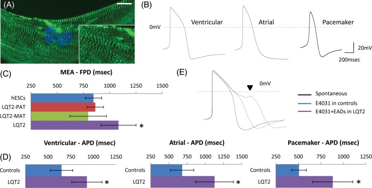
Long-QT syndrome-type 2 hiPSC cardiomyocytes can recapitulate the disease phenotype in vitro. (A) Long-QT syndrome-type 2 cardiomyocytes showed characteristic cardiac muscle striations with cardiac troponin-I staining, and (B) formed ventricular (n = 11), atrial (n = 14), and pacemaker-like (n = 9) myocytes. (C) Long-QT syndrome-type 2 cardiomyocytes had prolonged field potential duration (n = 10) and (D) action potential duration compared with controls, which included cardiomyocytes derived from human embryonic stem cells, the patient's father (long-QT syndrome-type 2-PAT) and mother (long-QT syndrome-type 2-MAT). (E) E4031 (1 µM) treatment caused prolongation in action potential duration of control cardiomyocytes (n = 6), depicting the presence of functional hERG channels, and caused arrhythmias only in long-QT syndrome-type 2 cardiomyocytes (n = 6).

