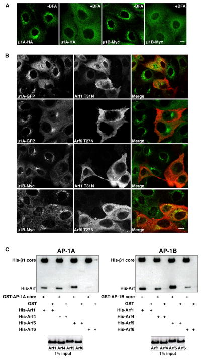
Figure 6. Similar Patterns of AP-1A and AP-1B Regulation by Arf Proteins.
(A) MDCK cells stably expressing μ1A-HA or μ1B-Myc were left untreated or treated with 5 μg/ml BFA for 15 min at 37°C, before immunostaining with antibodies to the HA or Myc epitopes. Scale bar, 10 μm.
(B) MDCK cells stably expressing μ1A-GFP or μ1B-Myc were transfected with plasmids encoding dominant-negative HA-tagged Arf1 T31N or Arf6 T27N mutants. At 24 hr after transfection, μ1A-GFP was detected by GFP fluorescence, and μ1B-Myc or Arf-HA was detected by immunostaining with antibodies to the Myc and HA epitopes. Scale bar, 10 μm.
(C) Purified GST-His-tagged AP-1A or AP-1B core complex was incubated with His-tagged Arf1, Arf4, Arf5, or Arf6 constitutively active (QL) mutants. Bound proteins were isolated on glutathione-Sepharose beads and analyzed by SDS-PAGE and immunoblotting with antibodies to the His tag. GST was used as a negative control.

