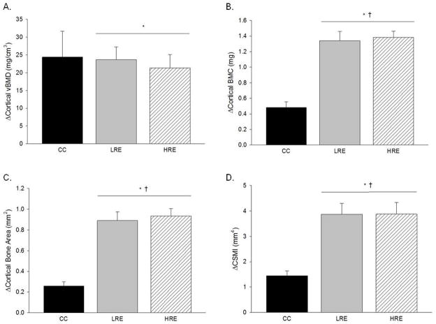
Figure 1.
Effects of low- and high-load resistance exercise (LRE, HRE) on changes in densitometric and geometric properties of the tibial mid-diaphysis as taken by in vivo pQCT scans. Changes are for the duration of the study (day 35 values minus day 0 values). A: Cortical volumetric bone mineral density (vBMD). B: Cortical bone mineral content (BMC). C: Cortical bone area. D: Polar cross-sectional moment of inertia (CSMI). Each bar represents the group mean ± standard error of the mean. † vs. CC (p ≤ 0.05); * vs. day 0 (p ≤ 0.05).

