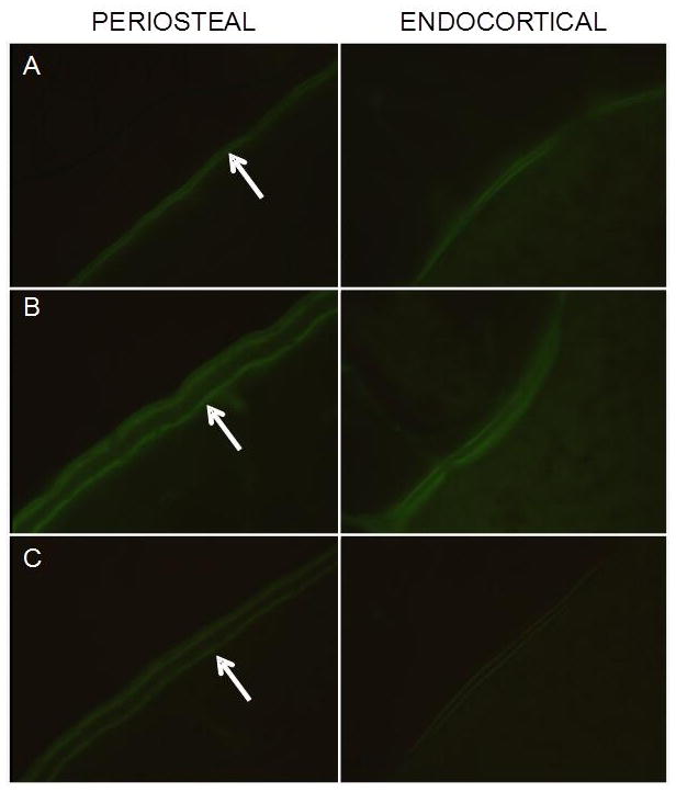
Figure 3.

Visual depiction (100x) of calcein labeling on the periosteal and endocortical surfaces of cortical bone at the tibia diaphysis. A: CC B: LRE C: HRE. Note the extensive flurochrome labeling (arrows) and large interlabel width (LRE and HRE).
