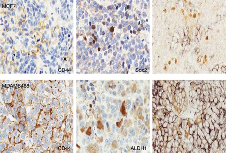
Figure 5.
Marker expression and co-localization in tumor xenografts. Immunocytochemistry for CD44 and Sox2 in MCF7 xenografts and for CD44 and ALDH1 in MDAMB468 xenografts as indicated. The image at the right-hand side of the two upper panels shows double staining of the two markers with CD44 in black and the other marker in brown. Single-label staining is counterstained with haematoxylin; no counterstain has been applied to double-labelled sections.

