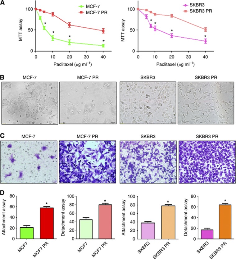
Figure 1.
PR cells exhibited EMT phenotype. (A) MTT assay was conducted in parental and PR breast cancer cells. *P<0.05 vs control. (B) Cell morphology was observed by microscopy in parental and PR cells. Parental MCF7 and SKBR3 cells displayed an epithelioid appearance, whereas their PR cells showed elongated, irregular fibroblastoid morphology. (C) Invasion assay was performed to measure the invasive capacity in parental and PR cells. (D) Cell attachment and attachment assays were assessed in parental and PR cells. *P<0.05 vs control.

