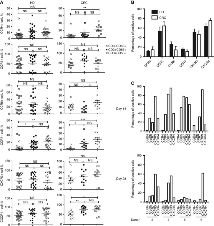
Figure 1.
CKR profiles on human CIK cell expansions. (A) Healthy donor (HD; n=28) and colorectal cancer patient (CRC; n=11) CIK cells at day 14 were stained for NK (CD56) and T-cell (CD3) markers, as well as the 6 chemokine receptors known to be expressed on CIK cells (CCR4, CCR5, CCR6, CCR7, CXCR3, CXCR4). Percentages of cells of different cell types (CD3−CD56+ CD3+CD56+ CD3+CD56−) positive for different CKRs are shown. (B) Comparisons of CKRs on CD3+CD56+ CIK cells from HD and CRC (no significant differences seen for any CKR; N.B. Other differences between these groups include average age (HD=39 years; CRC=53 years) and sex (HD=29% female; CRC=45% female). (C) Differences in CKR profiles in CD3+CD56+ CIK cell populations between donors (for 4 donors) and at different times after expansion are shown. Individual donors show distinct patterns of CKR profiles, which are gradually reduced over time in culture.

