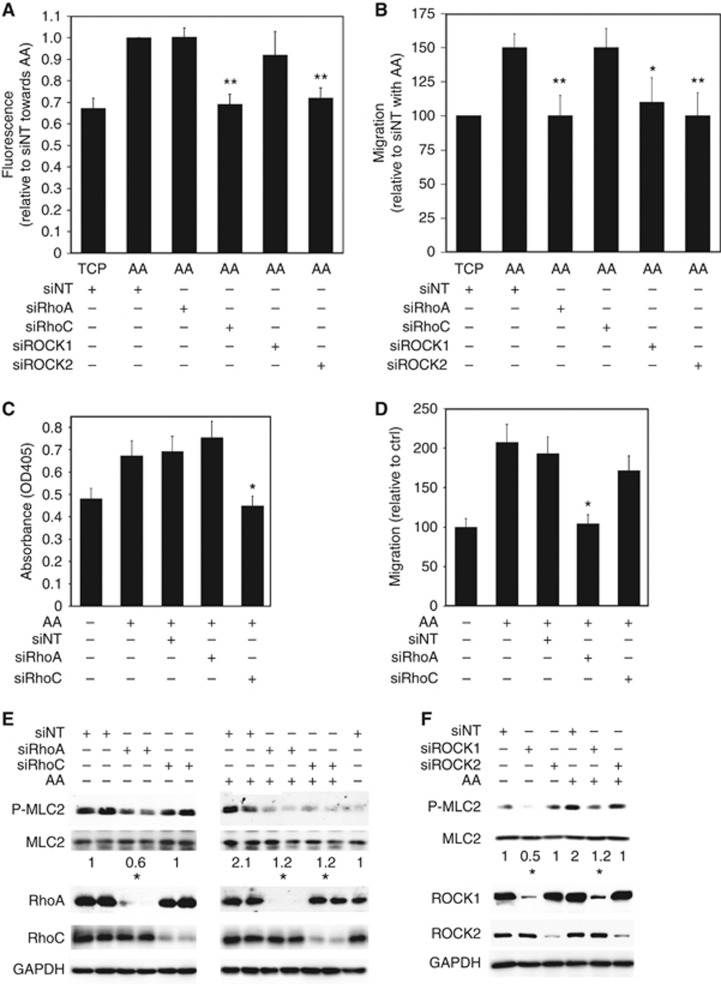
Figure 4.
AA-stimulated transendothelial migration is mediated by specific Rho GTPase signalling. (A) Adhesion assays were performed using PC3-GFP with or without AA (10 μM) added at the beginning of the assay. 4 × 10⁴ PC3-GFP cells were then incubated for 8 min with a confluent layer of BMEC plated in 96 wells. Wells were washed twice in PBS. Levels of adhesion are proportional to fluorescence detected by a bottom reading BMG FLUOstar OPTIMA plate reader at 488/520 nm (excitation/emission filter). PC3-GFP cells were transfected with specific RhoA (siRhoA), RhoC (siRhoC), ROCK1 (siROCK1), ROCK2 (siROCK2) siRNA pools, respectively, or with non-targeted siRNA (siNT) 2 days before the adhesion assays. Data represent means±s.e.m. of three separate experiments. **P⩽0.01 vs siNT with AA. (B) Migration assays were performed using PC3-GFP with or without AA (10 μM). PC3-GFP cells were transfected for 48 h with specific RhoA (siRhoA), RhoC (siRhoC), ROCK1 (siROCK1), ROCK2 (siROCK2) siRNA pools, respectively, or transfected with non-targeted siRNA (siNT) 48 h before the assay. Data represent means±s.e.m. of three separate experiments. *P⩽0.05, **P⩽0.01 vs siNT with AA. (C) Adhesion assays to BMEC over 15 min±AA (10 μM) were undertaken using DU-145 cells. These were transfected with specific RhoA (siRhoA) and RhoC (siRhoC) siRNA pools, respectively, or with non-targeted siRNA (siNT) 2 days before the adhesion assays. Data represent means±s.e.m. of three separate experiments. *P⩽0.05 vs no SIM with AA. (D) Migration assays were performed using DU-145±AA (10 μM). DU-145 cells were transfected with specific RhoA (siRhoA) and RhoC (siRhoC) siRNA pools, respectively, or with non-targeted siRNA (siNT) 2 days before the migration assays. Data represent means±s.e.m. of two separate experiments. *P⩽0.05 vs no SIM with AA. (E) P-MLC2t18/s19, MLC2, RhoA and RhoC content was analysed by western blotting in lysates of PC3-GFP cells transfected for 48 h with specific RhoA (siRhoA) and RhoC (siRhoC) siRNA pools, respectively, or transfected with non-targeted siRNA (siNT) and incubated with or without AA (10 μM) for 75 min. Figure is representative of three separate experiments. *P⩽0.05 vs siNT±incubation with AA. Densitometric values of siNT±incubation with AA were set at one. (F) P-MLC2t18/s19, MLC2, ROCK1 and ROCK2 content was analysed by western blotting in lysates of PC3-GFP cells transfected for 48 h with specific ROCK1 (siROCK1) and ROCK2 (siROCK2) siRNA pools, respectively, or transfected with non-targeted siRNA (siNT) and incubated±AA (10 μM) for 75 min. Figure is representative of three separate experiments. *P⩽0.05 vs siNT without or with incubation with AA. Densitometric values of siNT with or without incubation with AA were set at one.

