Fig. 1.
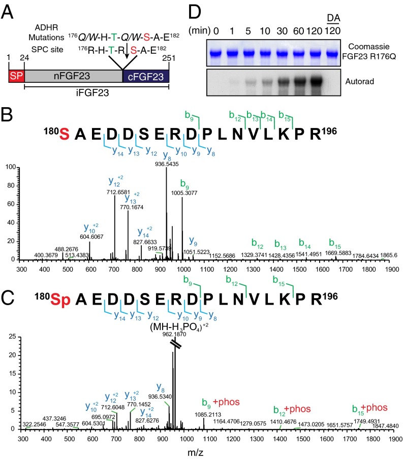
Fam20C phosphorylates FGF23 on Ser180. (A) Schematic representation of human FGF23 indicating the signal peptide (SP), intact FGF23 (iFGF23), N-terminal fragment (nFGF23), and C-terminal fragment (cFGF23). The residues surrounding the SPC cleavage site (downward arrow) are shown. Mutations that replace the Arg in patients with autosomal dominant hypophosphatemic rickets (ADHR) are also shown. A potential Fam20C phosphorylation site is in red. (B and C) Representative MS/MS fragmentation spectra of a tryptic peptide (FGF23 180–196) depicting Ser180 phosphorylation of FGF23 R176Q purified from conditioned medium of HEK293T cells. (D) Time-dependent incorporation of 32P from [γ-32P]ATP into FGF23 R176Q by Fam20C or Fam20C D478A (DA). Reaction products were analyzed by SDS/PAGE and autoradiography.

