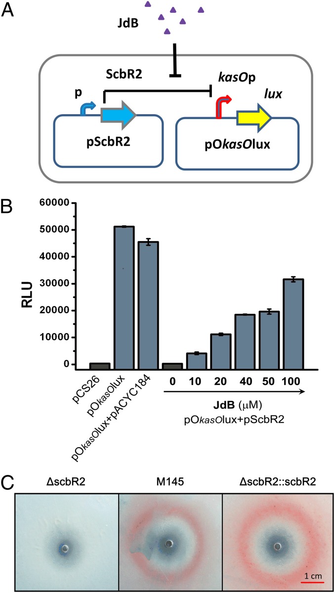
Fig. 3.
Demonstration of the response of ScbR2 to JdB. (A) A schematic representation of the reporter system used to investigate the response of ScbR2 to JdB. (B) Investigation of the interactions of JdB with ScbR2 using a Lux reporter system in vivo. pCS26 contains a promoterless lux operon, and pACYC184 was used to express scbR2. They were used as controls. Relative light units (RLU) are represented as the average of at least three independent readings; error bars indicate ±SDs. (C) Plate assay of the responses of ΔscbR2 (Left), M145 (Center), and ΔscbR2::scbR2 (Right) to JdB on SMM agar. JdB was dissolved in DMSO, and 10 μL (10 mM) was spotted onto a lawn of S. coelicolor. The photographs were from the bottom of the plates.

