Fig. 3.
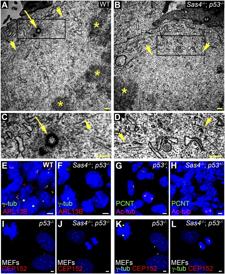
Sas4−/− embryos lack centrioles, centrosomes, and cilia. (A–D) TEM of E8.5 neural folds. Microtubules (arrowheads) converge at metaphase spindle poles, where there are centrioles (arrow) in wild-type but not Sas4−/− p53−/− embryos. Asterisks mark the condensed chromosomes in A and B. C and D are magnifications of the boxed regions at the spindle poles in A and B, respectively. (E–H) Expression of the centrosomal and cilia markers in the mesenchymal cells in E8.5 wild-type and Sas4−/− embryos stained for γ-tubulin (γ-tub) or PCNT (green) and ARL13B (red) or Ac-tub (magenta). Cilia are absent, and focal staining of γ-tubulin and PCNT is detected only during mitosis in Sas4−/− mutants. (I–L) Expression of the PCM proteins CEP152 (red) and γ-tubulin (green) in p53−/− MEFs [45 ± 5 arbitrary units (A.U.)] and Sas4−/− p53−/− MEFs (25 ± 9 A.U., P < 0.0001, n = 14 poles each). CEP152 is not detected in either interphase or mitotic cells in Sas4−/− mutants. (Scale bars: 0.3 µm in A–D, 3 µm in E–L.)

