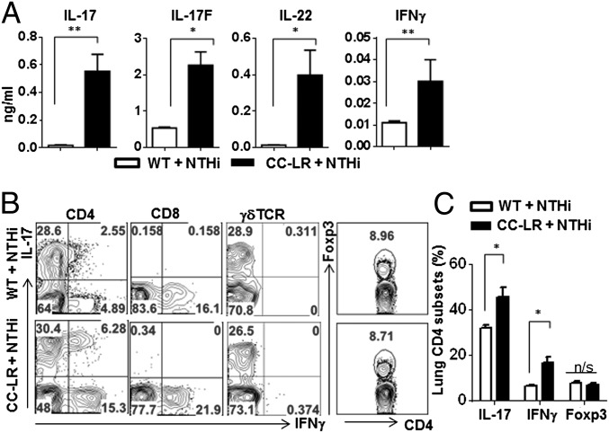
Fig. 2.
Increased Th17 cells in inflammation-promoted lung cancer. (A) Cytokines in BALF by ELISA (n ≥ 5 per group). (B) Representative flow cytometry plots of T cells. Lung mononuclear cells were isolated from 14-wk-old WT or CC-LR mice which were challenged with NTHi lysate for 4 wk. Isolated cells were stimulated with PMA/Ionomycin and stained with antibodies to CD4, CD8, γδTCR, IL-17, IFN-γ, and Foxp3. (C) Frequency of lung CD4 T cells expressing IL-17, IFN-γ, and Foxp3. Data are shown as mean ± SEM, *P < 0.05, **P < 0.01 (WT + NTHi, n = 6; CC-LR + NTHi, n = 8).

