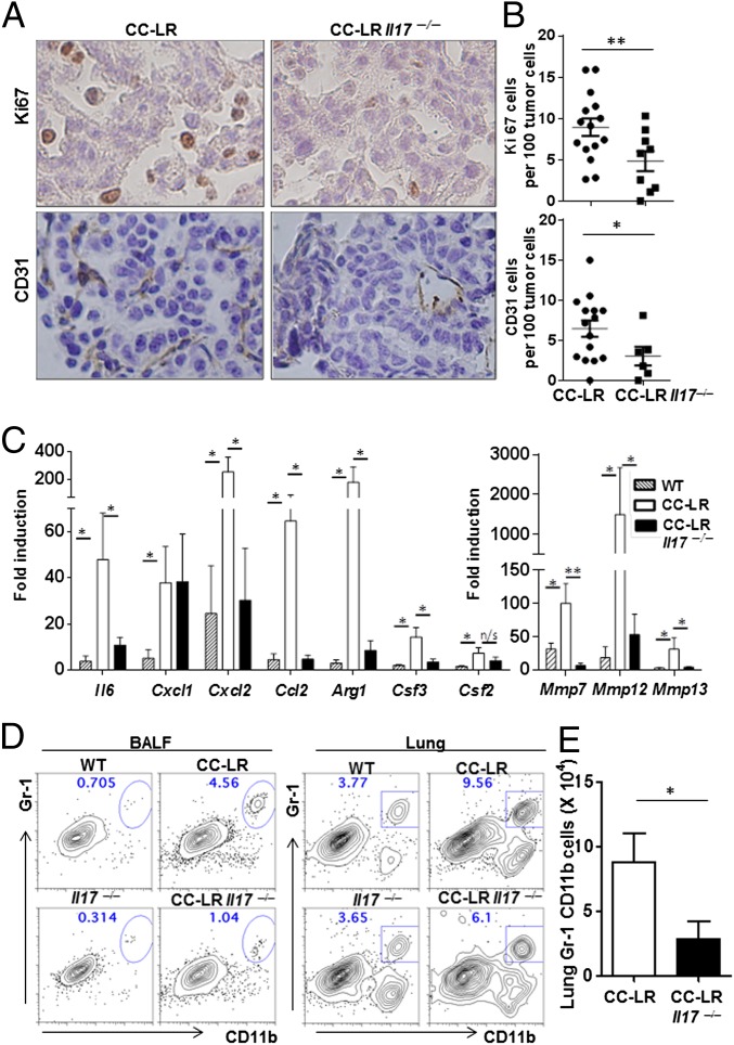
Fig. 4.
IL-17 deficiency reduces inflammation and cancer growth. (A) Representative immunohistochemistry of Ki67 and CD31 staining. (B) Ki67-positive cells or CD31 positive cells per 100 tumor cells at the 14-wk time point. n = 4 per group, 3–7 fields per mouse. (C) Relative expression of mRNA in whole lungs in 14-wk-old CC-LR mice (WT, n = 6; CC-LR Il17+/+, n = 6; CC-LR Il17−/−, n = 5). Data are expressed as fold increase compared with controls. Data represent means ± SEM *P < 0.05. **P < 0.01. (D) Representative flow cytometric plots of BALF or lung cells from WT and tumor-bearing mice. Cells were stained with anti-CD11b and Gr-1 Abs and gated in FSChi SSChi. (E) Absolute number of Gr-1+CD11b+ cells. Data are shown as means ± SEM (n = 5 per group, *P < 0.05).

