Fig. 1.
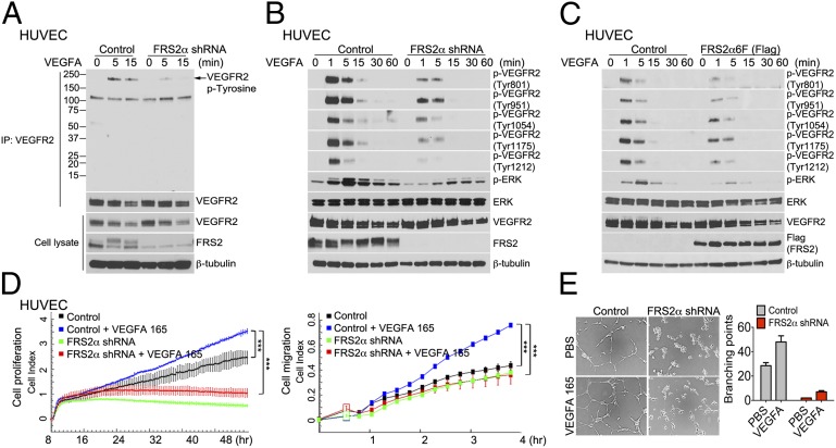
FRS2α knockdown in HUVEC inhibits VEGF-A165–dependent signaling. (A) Control and FRS2α knockdown HUVEC were serum starved overnight and treated with VEGF-A165 (50 ng/mL) as indicated. Cell lysates were immunoprecipitated (IP) with an anti-VEGFR2 antibody and immunoblotted with anti–p-Tyrosine antibody. The same blot was stripped and blotted with anti-VEGFR2. Input lysates were blotted with anti-VEGFR2 and anti-FRS2α antibodies. (B) Control and FRS2α knockdown HUVEC were serum starved overnight and treated with VEGF-A165 (50 ng/mL) as indicated. Cell lysates were blotted with p-VEGFR2, VEGFR2, p-ERK, ERK, and FRS2α antibodies. (C) Control and FRS2α-6F (Flag) overexpressed HUVEC were serum starved overnight and treated with VEGF-A165 (50 ng/mL). Cell lysates were blotted with p-VEGFR2, VEGFR2, p-ERK, ERK, and Flag (FRS2α) antibodies. (D) Control and FRS2α knockdown HUVEC were serum starved overnight. Cell proliferation (Left) and cell migration (Right) in response to VEGF-A165 (50 ng/mL) profiles are shown as detected by xCELLigence. Approximately 1000 cells (for cell proliferation) or 25,000 cells (for cell migration) were loaded per well in duplicate (***P < 0.01 compared with control). (E) In vitro Matrigel: The extent of cords branching was assessed in control and FRS2α knockdown HUVEC placed on growth factor-depleted Matrigel and exposed to VEGF-A165 (50 ng/mL). Data in A–C and E are based three independent experiments; data in D are based on two independent experiments.

