
Fig. 1.
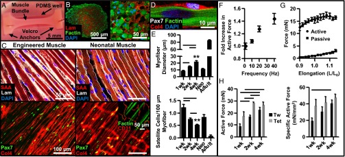
Structural and functional characterization of in vitro-engineered skeletal muscle. (A) Live image of a 2-wk engineered muscle bundle (∼1.5-mm diameter, 1.25 cm long) anchored at ends by tendon-mimetic Velcro tabs pinned inside a polydimethylsiloxane (PDMS) well. (B) Immunostained bundle cross-section shows F-actin+ myofibers embedded within laminin (Lam)-rich matrix. (C) Structural organization of representative engineered and native neonatal rat soleus muscles. (Inset) Transverse Col4+ structures present in native but not engineered muscle are CD31+ blood vessels. Col4, collagen IV; SAA, sarcomeric α-actin. (D) Pax7+ satellite cells in engineered muscle reside at myofiber sarcolemma. (E) Average myofiber diameter and SC number per 100-µm myofiber length at 1, 2, and 4 wk of culture compared with native neonatal (Neo) and adult soleus muscles. (F) Dependence of active force amplitude (normalized to that of single twitch) on stimulus frequency. (G) Dependence of active twitch and passive tension amplitudes on engineered muscle length (expressed relative to culture length). (H) Absolute and specific (force per area) twitch (Tw) and tetanus (Tet, 40 Hz) amplitudes in engineered bundles at 1, 2, and 4 wk of culture. Mean ± SEM; n = 4–10 samples per group (8–10 images per sample); **P < 0.01 between 4-wk bundle and native muscles; P < 0.05 between denoted groups.