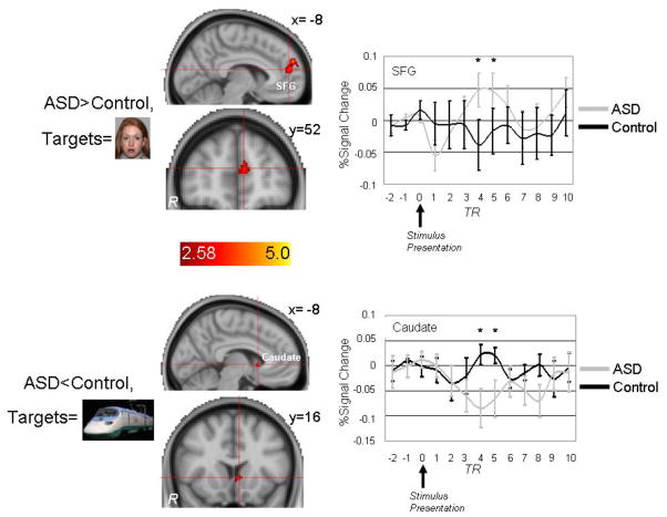
Figure 3.
Top: Brain areas showing increased activation to face targets in ASD participants relative to controls included two superior frontal gyrus (SFG) clusters and a cluster within right insular cortex (not shown). Bottom: Brain areas showing decreased activation to High Autism Interest (“HAI”) targets in ASD participants relative to controls included the left caudate nucleus. p<.05; Coordinates are in MNI space.

